日期:March 2021
作者:erin trachet,bs |科学发展助理主任
Lung cancer frequently metastasizes to other parts of the body. One of the most dangerous areas it can travel to is the brain, significantly reducing life expectancy. Unfortunately, these metastases are common. Up to 7% of people already have cancer cells in the brain when they are first diagnosed with non-small cell lung cancer (NSCLC), and 20% to 40% of those with NSCLC will develop this complication during disease progression. Brain metastases occur in stage 4 lung cancer. Stage 4 lung cancer has a poor prognosis, with life expectancy usually being under a year1. Currently there are no targeted therapies specific for brain metastases, and the blood-brain barrier can pose a physiologic impediment to many cytotoxic drugs and antibody-based therapies. Surgical tumor resection and/or radiotherapy are the most used forms of treatment, but the overall increase in survival is small (~4-6 months) and the quality of life following the treatment can be very poor2. Clearly, more effective treatments are needed for this devastating disease. The literature reveals very few preclinical oncology models that facilitate the assessment of metastatic brain disease in the presence of a primary tumor. In recognition of this need, Preclinical Oncology (PCO) at Covance has developed two xenograft models to not only address metastatic brain disease via a direct intra-cranial implant, but also couples this with a subcutaneous “primary” tumor to effectively allow the evaluation of the response to treatment at both locations.
此外,能够在两个植入物位置同时评估靶向疗法的药代动力学和/或药效学(PK / PD)是重要的。靶向疗法的治疗可具有显着不同的渗透和吸收率,这将改变在任一组织的PK / PD评估。鉴于大脑中的独特环境和血脑屏障在两种位置比较的障碍物可以是评估治疗对成熟的转移性疾病和原发性肿瘤的影响的强大工具。
PCO优化了两种人类非小细胞肺癌细胞系的双疾病诱导参数;NCI-H1975-LUC和PC-9-LUC。两种细胞系已经用荧光素酶转染,以允许生物发光成像(BLI)来监测植入内部植入细胞的疾病进展。皮下“初级”肿瘤被屏蔽以防止重叠信号。在所有涉及动物的研究中,协议是根据AAALAC认可的设施的动物福利法规进行了IACUC议定书审查和批准。
NCI-H1975-Luc
NCI-H1975-Luc was established from a female non-smoker. This cell line is of interest to the research community because of its EGFR-L858R/T790M mutational status. The T790M acquired mutation is found in 50-60% of NSCLC patients that become resistant to approved EGFR inhibitors. Therefore, this model is suitable for evaluating next generation EGFR inhibitors, such as compounds that bind to EGFR regardless of the mutational changes that have occurred, or compounds that bind irreversibly. The EGFR expression profile of NCI-H1975-luc has made it a model of interest for the dual implant method because it allows for the assessment of blood brain barrier penetration of an EGFR targeted small molecule. This evaluation can be done multiple ways; traditional efficacy based on BLI monitoring of the metastatic brain disease coupled with caliper measurements of the subcutaneous tumors and/or pharmacodynamic and pharmacokinetic analysis of the metastatic lesion compared to the primary tumors to assess if drug concentration is different. In either scenario the metastatic brain disease response can then be compared to the “primary” subcutaneous tumor response.
与该模型的历史肿瘤生长一致,皮下肿瘤体积倍增时间约为7天,通常达到评价大小(〜900mm3) in approximately 25 days post implant (Figure 2). As expected, the model is just as reliable when implanted intra-cranially, with a BLI calculated tumor volume doubling time of 1.6 days (Figure 1 and Image 1).
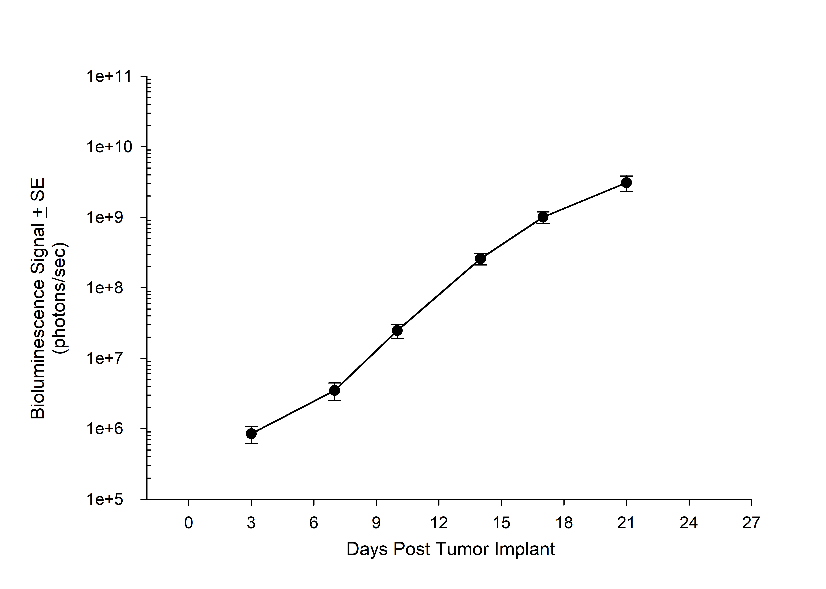
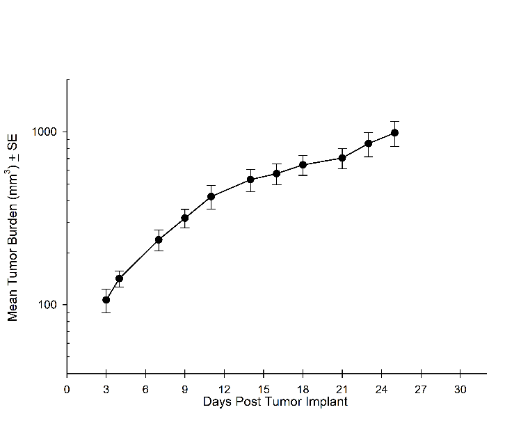
Figure 1A: NCI-H1975-Luc Mean BLI Signal for Metastatic Disease Progression
图2A:NCI-H1975-LUC意味着初级(SC)肿瘤生长
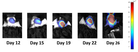
Image 1: NCI-H1975-Luc Representative Images of Metastatic Brain Disease Progression
PC-9-LUC
1989年,从雄性肺腺癌患者中分离PC-9-LUC。据报道,PC-9-LUC对吉替尼和其他EGFR酪氨酸激酶抑制剂非常敏感。然而,已经表明,将PC-9-LUC细胞的延长暴露于EGFR抑制剂可能导致采集T790M突变和抗性细胞系。当评估T790M突变的演变,用于预防突变的潜在治疗或建立抗性细胞系后处理时,PC-9-LUC模型是有价值的。类似于NCI-H1975-LUC细胞系,Covance开发了双植入参数,其允许评估原发性肿瘤和转移性脑疾病。
Consistent with the historical tumor growth for this model, the subcutaneous tumor volume doubling time is about 4 days and typically reaches evaluation size (~1000mm3) in approximately 20 days post implant (Figure 4). As expected, the model is just as reliable when implanted intra-cranially, with a BLI calculated tumor volume doubling time 2.6 days (Figure 3 and Image 2).
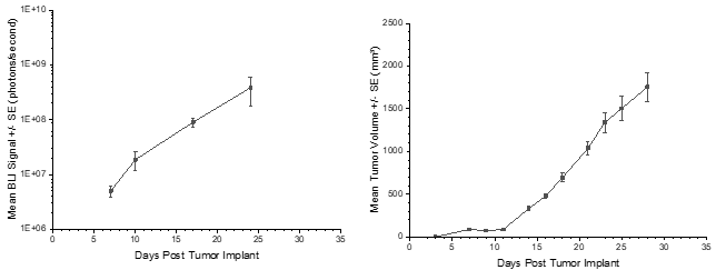
Figure 3: PC-9-Luc Mean BLI Signal for Metastatic Disease Progression
图4:PC-9-LUC意味着初级(SC)肿瘤生长
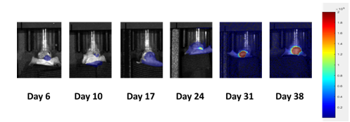
Image 2: PC-9-Luc Representative Images of Metastatic Brain Disease Progression
ContactCovance的科学家们要求完整的细胞系列列表或了解有关如何与我们的光学成像服务相结合的皮下,转移和原位模型的更多信息,可以应用于您的临床前研究。yaboapp体育官网
References
1Ho Jeong Lee,† Masaki Hanibuchi,† Sun-Jin Kim,† Hyunkyung Yu, Mark Seungwook Kim, Junqin He, Robert R. Langley, François Lehembre, Urs Regenass, and Isaiah J. Fidler. Treatment of experimental human breast cancer and lung cancer brain metastases in mice by macitentan, a dual antagonist of endothelin receptors, combined with paclitaxel. Neuro Oncol. 2016 Apr; 18(4): 486–496.
2Cairncross JG,Kim JH,Posner JB。脑转移的放射疗法。Ann Neurol。1980; 7(6):529-541。
Note: Please note that allanimal care and usewas conducted according to animal welfare regulations in an AAALAC-accredited facility with IACUC protocol review and approval.
Let's start a conversation
联系我们
景by Labcorp is a leading global life sciences company, which provides contract research services to the drug, medical device and diagnostics, crop protection and chemical industries.
Covance是Covance Inc.的注册商标和营销名称及其周围的子公司。