Lung cancer is the most common cause of cancer related deaths for men and second for women (after breast cancer). Worldwide, 1.8 million diagnoses and 1.6 million deaths occur annually.1虽然近几十年来的事件一直在减少,但诊断和治疗的进步是整体癌症,只有17%的患者患有肺癌的患者将存活五年,为新的疗法提供了对这一人口的巨大影响的机会患者。手术,传统化学疗法,靶向疗法,辐射和免疫疗法的组合已被证明是有益的治疗选择;并开发新颖的疗法和/或组合继续。
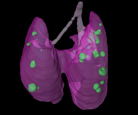
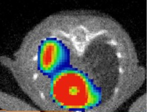
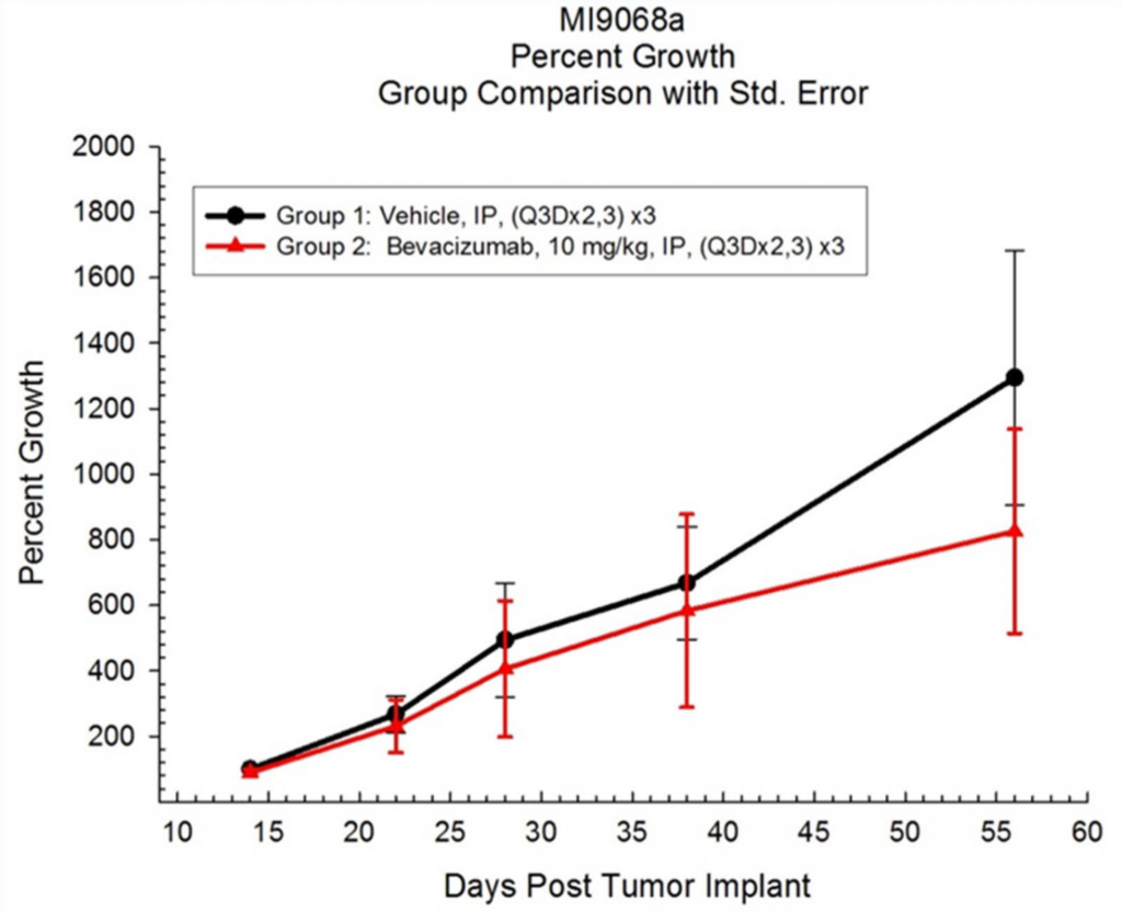
Clinical imaging technologies have been vital for detecting and following disease progression/regression for many years, as examples, Computed Tomography (CT) can measure tumor volume over time and Positron Emission Tomography (PET) can measure metabolic activity within the tumors. At Covance we have been utilizing preclinical imaging technologies for over a decade to evaluate tumors in the lung from orthotopic implants of syngeneic, primary tumor xenografts (PDX), GEM models and metastatic disease (Figures 1, 2, and 3). Now with the increase of interest in immunotherapies, where the extracellular matrix can play a significant role in the engagement of immune cells, the stroma now is of greater importance;2, 3使用成像允许治疗评估在适当的基质中。
Covance具有将正确的成像技术应用于适当的肿瘤模型,使您的研究施加。Contact Covance有关详细信息。