日期:2021年5月
联系临床前肿瘤科学团队以了解更多信息。
Covance by Labcorp Preclinical Oncology offers the new mouse Expanded CompMyeloid™ panel, which provides deep immunophenotypic analysis into myeloid subsets known to regulate the tumor immune response. The panel addresses the growing need to understand how immunotherapy modulates pro-tumor and anti-tumor activity in the myeloid compartment of the tumor microenvironment. In this Tech Spotlight, we demonstrate the power of this 18-color panel that provides a deep dive into myeloid subset immunophenotyping and functional characterization.
The Expanded CompMyeloid™ panel builds upon our CompMyeloid™ panel, which provides a general measurement of five myeloid subsets that regulate immunity in the tumor. The improved panel adds DC1 (type 1) and DC2 (type 2) dendritic cell delineation, plus six additional markers for functional characterization of tumor-associated macrophage (TAM), myeloid-derived suppressor cell (MDSC), and dendritic cell subsets. Table 1 describes the components of the Expanded CompMyeloid™ panel and Figure 1 illustrates the gating strategy for myeloid subset delineation using the murine Hepa 1-6 hepatocellular carcinoma model.
Table 1: Expanded CompMyeloid™ Panel Antibodies and Description of Their Utility
繁体/染料 |
描述 |
CD45 |
泛免疫细胞标记物 |
CD11B. |
Pan-myeloid lineage marker/DC2 subset marker |
CD103 |
DC1 subset marker |
Ly-6G |
Granulocytic-MDSC/ Neutrophil marker |
Ly-6C |
单核细胞 - MDSC /单核细胞标记 |
CD11C. |
树突状细胞标记物 |
CD24 |
Dendritic cell delineation |
F4/80 |
泛巨噬细胞标记 |
MHC II类 |
M1巨噬细胞和树突状细胞标记 |
CD206 |
M2 macrophage marker |
XCR1 |
交叉呈递/迁移树突细胞标记 |
CD80 |
成熟标记物 |
CD86 |
成熟标记物 |
ARG1 |
骨髓功能标记 |
伊克斯 |
骨髓功能标记 |
PD-L1 |
T cell inhibitory signaling protein |
CD19 |
B cell marker |
活性染料 |
死细胞排除 |
The Expanded CompMyeloid™ panel provides immunophenotypic analysis of 7 distinct myeloid subsets including G-MDSC/neutrophils, M-MDSC/monocyte subsets, M1 and M2 Macrophages, and DC1, DC2 and XCR1+ dendritic cell (DC) subsets. The analysis of 5 myeloid functional markers to profile dendritic cell, macrophage, and MDSC phenotypes is also provided.
|
|
Expanded CompMyeloid™ Panel Gating Strategy
在Labcorp临床前肿瘤学免疫脑型面板与所有Covance对齐,分析以死区排除和yaboapp体育官网随后的CD45开始+immune cell delineation. Gated on total CD11b+cells, Figure 1A displays downstream MDSC and TAM analysis. Figure 1B demonstrates DC analysis that follows a meticulous gating strategy that first excludes macrophages, granulocytes, and B cells, which minimizes the risk that the DC measurement is compromised due to contamination by irrelevant and potentially autofluorescent cells. The exclusion gate then enables CD24 analysis (not shown) that helps delineate conventional DCs, which are generally classified by medium to high expression levels of CD11c and MHC class II. These conventional DCs include the CD103+DC1子集和CD11b+DC2 subset, which have been demonstrated to drive CD8+and CD4+T细胞抗肿瘤反应1。XCR1 expression has been documented by multiple groups as a marker for DCs with cross-presenting activity, a phenotype required for DC-mediated activation of CD8+T cells2。
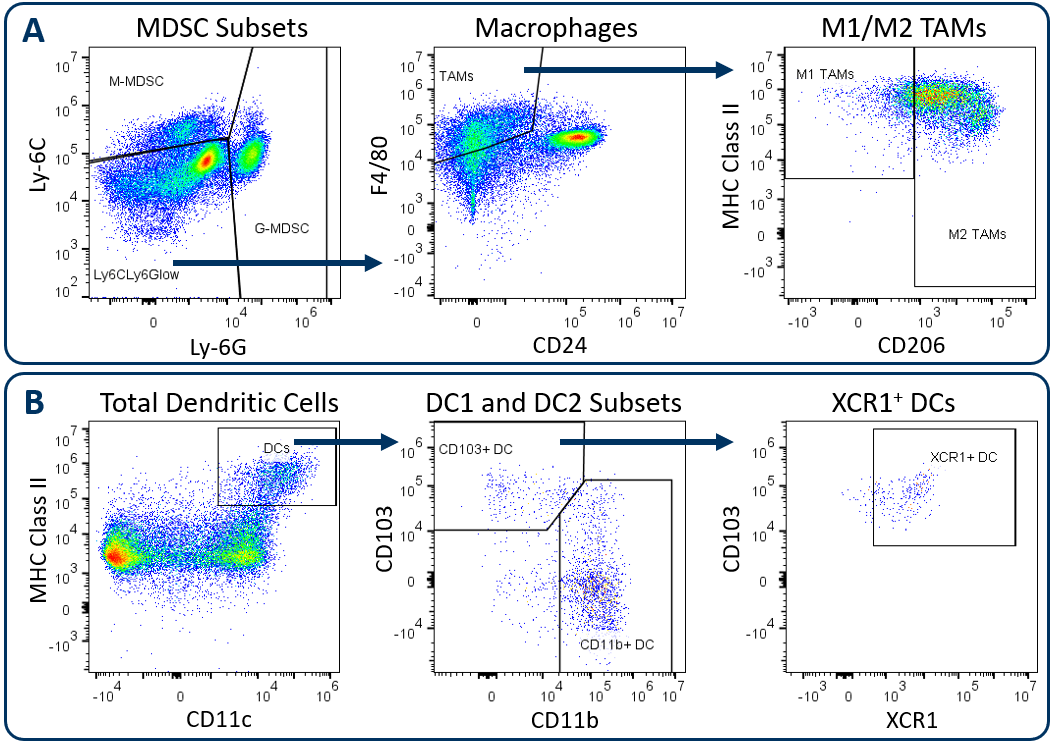
图。1。Analysis of tumor-infiltrating myeloid subsets using the Expanded CompMyeloid™ panel. Hepa 1-6 tumors were harvested from C57BL/6 mice. (A) Starting from a CD11b+cell gate, G-MDSC and M-MDSC subsets are measured and the remaining events are delineated into M1 and M2 TAMs, (B) Starting from a macrophage/MDSC exclusion gate, DC1 and DC2 subsets, and an XCR1+测量DC分数。
All animal work is approved by the site Institutional Animal Care and Use Committee and was performed in conformance with theGuide for the Care and Use of Laboratory Animalswithin an AAALAC-accredited program. Humane euthanasia criteria are predetermined on all studies.
伊克斯and Arginase 1 Analysis in MDSC and TAM Subsets
骨髓亚群赋予肿瘤微环境的活性,所述肿瘤微环境具有亲肿瘤和抗肿瘤作用。扩展的Compmyeloid™面板提供INOS和Aginase 1(Arg1)的分析;是髓样功能的二元性的实例的生物标志物。INOS和ARG1在肿瘤中的MDSC和TAM子集中表达3。在MDSCS中,INOS和ARG1通过竞争燃料T细胞增殖和抗肿瘤反应的基材L-精氨酸竞争免疫抑制效果。具有高InOS和Arg1表达的肿瘤可导致L-精氨酸剥夺的环境导致差的T细胞活化。因此,减少MDSC中InOS和Arg1表达的疗法具有促进T细胞活化和改善临床结果的可能性。值得注意的是,INOS表达还可以抑制T细胞无关方式的肿瘤生长,因为一氧化氮基因产物可以对肿瘤细胞具有直接细胞毒性作用以抑制疾病进展。由M1偏振巨噬细胞表达的INOS可以诱导生长逮捕以及在不同模型中诱导凋亡细胞死亡4,5。相反__arg1 expression by M2 polarized macrophages in the tumor microenvironment suppress T cell responses in a manner mechanistically similar to MDSCs.
Figure 2 demonstrates how the Expanded CompMyeloid™ panel can examine iNOS and ARG1 in myeloid subsets, using the subcutaneous Hepa 1-6 hepatocellular carcinoma model. We used this model because checkpoint inhibition via anti-PD-1 treatment inhibits tumor growth (not shown), thus providing an opportunity to examine how the myeloid compartment responds during checkpoint blockade. Compared to control animals, anti-PD-1 treatment increased the expression of both iNOS and ARG1 in tumor-infiltrating granulocytic (G-) and monocytic (M-) MDSC subsets (Figure 2A). In contrast, analysis of total TAMs demonstrated an increase in iNOS expression coinciding with a small but significant reduction in ARG1 expression (Figure 2B). Notably, within iNOS positive TAMs, there appear to be cells with a bi-phasic iNOS expression profile. Moreover, the expression of MHC class II is higher in the iNOS高的细胞级分,表明抗原呈递和T细胞激活的增强能力。作为M1和M2偏振的标记分别是INOS和ARG1,数据表明,抗PD-1治疗将TAM平衡移向M1偏振和抗肿瘤表型。数据也提醒肿瘤免疫反应是动态的,并且这种免疫疗法可以引发推动和拉动的多层效果,然后在肿瘤和抗肿瘤活性之间拉动。

Figure 2. iNOS and arginase 1 analysis in MDSC and TAM subsets. The Expanded CompMyeloid™ panel measures biomarkers for myeloid functionality. In this data, Hepa 1-6 tumors were harvested from anti-PD-1 and isotype control antibody treated mice. MDSCs (A) and TAMs (B) were then delineated and analyzed for iNOS and ARG1 expression. TAMs differentially expressed relatively high (red circle enclosed) and low (blue circle enclosed) levels of iNOS expression, which directly correlated with MHC class II expression in these cell fractions, which is consistent with M1 polarization (red and green gates are representative examples and were not in this case used to generate the MHC class II measurement shown). * Student's T Test (p<0.05).
CD80 and CD86 Maturation Marker Expression on Conventional DC Subsets
Conventional dendritic cells at work in the tumor microenvironment can be divided predominantly into two subsets called DC1 and DC2, which are identified by expression of CD103/XCR1 and CD11b respectively. Both subsets can activate T cells. However, DC1 cells have been shown to favor CD8+T细胞活化由于肿瘤相关抗原在排水淋巴结中的交叉呈递的增强容量,由XCR1表达调节的过程6。In the Hepa 1-6 study described above, the Expanded CompMyeloid™ panel was used to measure CD80 and CD86, which are costimulatory molecules expressed on antigen presenting cells and required for maximum T cell activation through a cell contact-dependent mechanism7。Figure 3 illustrates that both CD80 and CD86 are detectable on Hepa 1-6 tumor-derived dendritic cells. Furthermore, treatment with anti-PD-1 increased the expression levels of CD86 on the DC1 subsets compared to the isotype control group. The data suggest that enhanced T cell activation may be triggered by anti-PD-1 antibody treatment that is in part due to increased expression of CD86 on DC1 cells to enhance costimulatory activity in this subset.
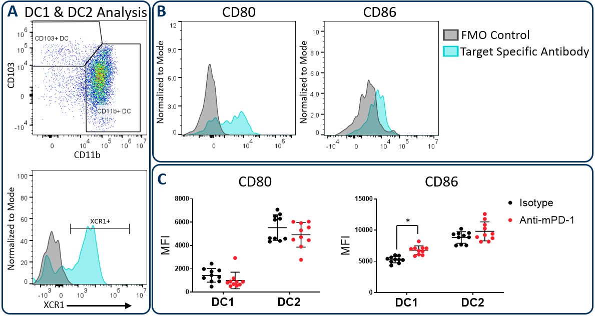
Figure 3. CD80 and CD86 maturation marker expression on DC subsets. The Expanded CompMyeloid™ panel was used to measure CD80 and CD86 in Hepa 1-6 tumor-derived cells. (A) Total DCs were first delineated and then phenotyped for DC1 and DC2 subsets. The gating strategy for XCR1 measurement is also shown. (B) Histograms to demonstrate representative CD80 and CD86 signals compared to the fluorescence minus one (FMO) negative control in DC1 cells. (C) CD80 and CD86 was measured in DC1 and DC2 subsets from Hepa 1-6 tumor-derived cells. * Student's T Test (p<0.05).
PD-L1 Analysis on Myeloid Subsets and Tumor cells
PD-1 / PD-L1轴是癌症免疫疗法中最广泛的靶向途径之一。PD-L1可以在肿瘤细胞上上调作为逃避宿主保护性免疫的机制。此外,已经记录了PD-L1表达,在粘蛋白细胞上,包括巨噬细胞,树突细胞和MDSC,以进一步抑制T细胞活化8。在一些情况下,这些独特的细胞类型中的PD-L1表达可以具有非冗余角色9。这些观察结果强调了了解肿瘤骨髓室中PD-L1表达的重要性可以通过免疫疗法调节。图4展示了扩展的CompMyeloid™面板在总TAM,DC,G-MDSC / M-MDSC子集以及CD45上测量PD-L1的能力-(非免疫)细胞,包括肿瘤细胞,以及血管内皮和其他细胞。如下所示,在HEPA 1-6肿瘤衍生细胞中检查的所有细胞类型中可检测到PD-L1。数据还表明,与对照动物相比,抗PD-1治疗对这些细胞中的PD-L1表达水平没有影响,表明PD-L1未通过该模型中的检查点抑制调节。
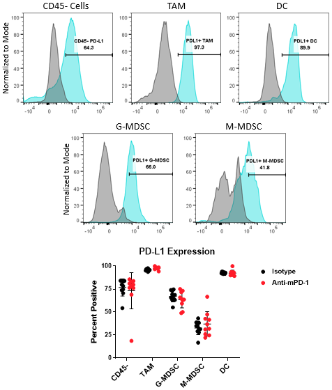
Figure 4. PD-L1 analysis on myeloid subsets and tumor cells. Histograms demonstrate representative PD-L1 signals compared to the fluorescence minus one (FMO) negative controls in CD45-来自HEPA 1-6肿瘤衍生细胞的细胞和骨髓亚群。所示的门用于量化在同种型对照和抗PD-1抗体处理基团中所示的所指示的子集中的PD-L1表达阳性的细胞的百分比。
通过使用扩展的Compmyeloid™面板,该技术聚焦的数据呈现了检查点抑制在鼠HEPA 1-6肿瘤模型中的多个骨髓亚群中的免疫活性调节。此外,数据提供了额外的支持,即抗PD1处理施加不受仅限于T细胞活动的效果。要了解有关扩展Compmyeloid™面板如何合并到您的临界研究中,请通过Labcorp与Covance的科学家联系。yaboapp体育官网
参考资料
1Gardner, A.和Ruffell, B. (2016)。树突状细胞与癌症免疫。免疫学趋势,37(12),855-865。
2Wylie,B.,Seppanen,E.,Xiao,K.,Zemek,R.,Zanker,D.,Prato,S.,...&Waithman,J.(2015)。迁移性XCR1+ CD103-和XCR1 + CD103 +树突状树突状细胞对皮肤皮肤的交叉呈递。onchoimmunology,4(8), e1019198.
3Kumar, V., Patel, S., Tcyganov, E., & Gabrilovich, D. I. (2016). The nature of myeloid-derived suppressor cells in the tumor microenvironment. Trends in immunology, 37(3), 208-220.
4Rahat,M.A.,&Hemmerlein,B。(2013)。巨噬细胞肿瘤细胞相互作用调节一氧化氮的功能。生理学的前沿,4,144。
5Vannini, F., Kashfi, K., & Nath, N. (2015). The dual role of iNOS in cancer. Redox biology, 6, 334-343.
6Wylie,B.,Seppanen,E.,Xiao,K.,Zemek,R.,Zanker,D.,Prato,S.,...&Waithman,J.(2015)。迁移性XCR1+ CD103-和XCR1 + CD103 +树突状树突状细胞对皮肤皮肤的交叉呈递。onchoimmunology,4(8), e1019198.
7Slavik, J. M., Hutchcroft, J. E., & Bierer, B. E. (1999). CD28/CTLA-4 and CD80/CD86 families. Immunologic research, 19(1), 1-24.
8Sun, C., Mezzadra, R., & Schumacher, T. N. (2018). Regulation and function of the PD-L1 checkpoint. Immunity, 48(3), 434-452.
9OH,S. A.,Wu,D. C.,张,J.,Navarro,A.,Xiong,H.,Cubas,R.,...&Mellman,I.(2020)。树突细胞的PD-L1表达是癌症中T细胞免疫的关键调节因子。自然癌,1(7),681-691。
让我们开始对话
联系我们
Labcorp旗下业主部门科学公司是一家人的全球生科学公司,致力于向向物,医疗器材和诊断,作物保护和化学行业提供合并研究服务。
科文斯是Covance Inc. 及其在世界各地的子公司的注册商标,也是其市场称谓。